Brain MRI Changes Are Not Neuron Counts
Key takeaways
- MRI does not directly count new neurons or synapses.
- Structural and functional imaging measure different properties.
- Clinical benefit requires appropriate outcome evidence, not just a changed scan.
Why a larger region is not a simple verdict
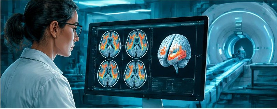
Reports that an activity makes a brain region larger attract attention because size sounds tangible. Structural MRI, however, measures signals that researchers use to estimate tissue properties and boundaries. It does not provide a direct inventory of neurons, synapses or useful memories. A measured volume difference can be scientifically important while leaving its exact cellular explanation uncertain.
Changes in tissue water, blood vessels, glial cells, myelination and other properties can affect imaging measurements. Processing choices and image quality also matter. The appropriate interpretation depends on the study, not a generic assumption that more gray matter always means better brain function. A smaller measurement is not automatically damage, and a larger one is not automatically improvement.
For further context, see a guide to reading brain-change research.
What the juggling study contributed
Draganski and colleagues' 2004 Nature report described gray-matter changes associated with learning to juggle in healthy adults. Its importance lies in demonstrating experience-associated structural imaging differences over a relatively short learning period. It supported the broader idea that the adult brain is not anatomically fixed in the simplistic sense often assumed.
The study was not a trial of dementia prevention, stroke rehabilitation or a general intelligence intervention. It does not justify prescribing juggling as treatment for those conditions. Nor does the finding identify a precise number of newly formed neurons. A durable reading preserves both parts of the result: the imaging observation matters, and the clinical claims it can support remain limited by the population, task and outcome.
For further context, see why blood BDNF is not a plasticity score.
Randomized exercise evidence adds a different piece
Erickson and colleagues studied aerobic exercise in older adults in a year-long randomized trial. The study reported increased anterior hippocampal volume in the aerobic-exercise group and memory-related findings. Randomization strengthens interpretation of differences between the assigned programs, but it does not transform MRI into a microscope or settle all mechanisms behind the observed volume change.
The trial supports discussion of a particular exercise intervention in its study population. It does not show that every workout produces the same effect, that an individual's scan can verify successful exercise or that hippocampal enlargement guarantees prevention of dementia. These broader claims require their own evidence. Health benefits of activity also need not depend on proving a visible brain change in each person.
Functional imaging answers another question
Functional MRI commonly tracks changes related to blood oxygenation, which are coupled indirectly to neural activity. Researchers can compare activation patterns or correlations between regions. These analyses are useful tools, but an altered connection on an imaging map is not a new anatomical cable. The word “connectivity” can refer to a statistical relationship rather than a direct structural link.
This matters across research areas. A small intensive human psilocybin imaging study reported altered synchronization patterns, for example, but that observation does not by itself establish treatment efficacy for depression or prove synapse growth. The same caution applies to training and rehabilitation imaging. Mechanistic findings can generate hypotheses about clinical benefit without replacing trials that measure symptoms, function and harms.
How convincing is the comparison?
Look for repeated measurements, an appropriate control group, transparent analysis and a clear relationship between the imaging result and the study question. Movement during scanning can distort findings, particularly when groups differ in their ability to remain still. Small samples and many analytic choices can create unstable results. Independent replication matters more than the apparent precision of a colorful image.
A correlation between brain change and improvement also does not prove that the measured change caused the improvement. Both may reflect another process, or the relationship may depend on a few participants. Ask whether the association was planned, whether it survives reasonable sensitivity checks and whether it predicts anything in a new sample. Those questions are more informative than simply asking which region lit up.
What this means for an individual reader
Suppose a wellness program offers a scan before and after training and promises proof of rewiring. Ask whether the measure is validated for individual decisions, whether its normal test-to-test variation is known and whether changing it improves an outcome you care about. A research measure that detects average group differences may be unsuitable for deciding whether one person's program worked.
Routine care should be guided by appropriate clinical assessment, not by a demand for an imaging trophy. Improved daily function can be meaningful without a follow-up scan. Conversely, a reported brain difference does not justify continuing an ineffective or burdensome program. The sensible stance is neither dismissal nor awe: MRI can reveal important patterns of adaptation, but its signals need careful interpretation and cannot carry every clinical claim attached to the word plasticity.
Sources
- Neuroplasticity: changes in grey matter induced by training.
- Erickson et al. (2011): Exercise training increases size of hippocampus and improves memory
- Psilocybin desynchronizes the human brain.
Editorial disclosure: It is educational information, not individual medical advice. Prepared for October 30, 2026 using established research; it does not assert current regulatory status or latest findings.
Written by
Neuroplasticity News Editorial Team
The Neuroplasticity News editorial team covers brain science, rehabilitation research and clinical trials, translating peer-reviewed evidence into clear, accurate reporting for patients, families and professionals.
 PreviousRehabilitation Progress: Measuring What Matters When Change Is Uneven
 NextConcussion Exercise Research: Why “Below the Symptom Threshold” Matters